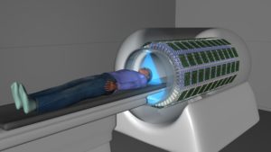
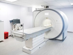
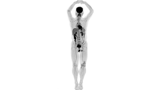
Created by researchers from UC Davis, the scanner consolidated positron emission tomography (PET) and X-beam processed tomography (CT) to catch the whole body without a moment’s delay.
Researchers Simon Cherry and Ramsey Badawi trust the innovation will have “incalculable applications” from enhancing diagnostics through to tranquilize research and following sickness movement.
The primary pictures of human sweeps from the EXPLORER are to be introduced at a gathering of the Radiological Society of North America on 24 November in Chicago.

“While I had envisioned what the pictures would look like for a considerable length of time, nothing set me up for the unimaginable detail we could see on that first sweep,” said Mr Cherry, recognized educator in the UC Davis bureau of biomedical building.
Mr Badawi, head of atomic drug at UC Davis Health and bad habit seat for research in the division of radiology, said he was puzzled when he saw the primary pictures.
“The dimension of detail was amazing, particularly once we got the reproduction technique more improved,” he said.

“We could see includes that you simply don’t see on customary PET sweeps. Also, the dynamic succession demonstrating the radiotracer moving around the body in three measurements after some time was, to be honest, awe-inspiring.”
Mr Badawi and Mr Cherry originally concocted the possibility of the full-body scanner over 10 years prior, however it wasn’t until 2011 that their thought was kick-begun with a $1.5m (£1.17m) concede from the US National Cancer Institute.
Another concede support in 2015, worth $15.5m (£12m), enabled them to expedite a business accomplice and really construct the principal EXPLORER unit.
Source: Sky News and New Atlas


